Daptomycin is a cyclic lipopeptide antibiotic used to treat complicated skin and skin structure infections by susceptible Gram-positive bacteria and bacteremia due to Staphylococcus aureus. Daptomycin is a cyclic lipopeptide antibacterial agent with a broad spectrum of activity against Gram-positive bacteria, including methicillin-susceptible and -resistant Staphylococcus aureus (MSSA/MRSA) and vancomycin-resistant Enterococci (VRE).
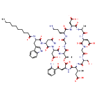
- Mechanism
- Curator reviewed · 24 references
The mechanism of action of daptomycin remains poorly understood. Studies have suggested a direct inhibition of cell membrane/cell wall constituent biosynthesis, including peptidoglycan, uridine diphosphate-N-acid, acetyl-L-alanine, and lipoteichoic acid (LTA). However, no convincing evidence has been presented for any of these models, and an effect on LTA biosynthesis has been ruled out by other studies in S. aureus and E. faecalis.
It is well understood that free daptomycin (apo-daptomycin) is a trianion at physiological pH, which binds Ca2+ in a 1:1 stoichiometric ratio to become a monoanion, which is thought to rely primarily on the Asp(7), Asp(9), and L-3MeGlu12 residues that form a DXDG motif. Calcium-binding facilitates daptomycin's insertion into bacterial membranes preferentially due to their high content of the acidic phospholipids phosphatidylglycerol (PG) and cardiolipin (CL), wherein it is proposed that daptomycin can bind two calcium equivalents and form oligomers. PG is recognized as the main membrane requirement for daptomycin activity; daptomycin preferentially localizes in PG-rich membrane domains, and mutations affecting PG prevalence are linked to daptomycin resistance. Calcium-dependent membrane binding is the generally accepted mechanism of action for daptomycin, but the precise downstream effects are unclear, and numerous models have been proposed.
One mechanism proposes that the daptomycin membrane binding alters membrane fluidity, causing dissociation of cell wall biosynthetic enzymes such as the lipid II synthase MurG and the phospholipid synthase PlsX. This is consistent with the observed effects of daptomycin on cell shape in various bacteria at concentrations at or above the minimum inhibitory concentration (MIC). Aberrant cell morphology is also consistent with the observed localization of daptomycin at the division septa and a hypothesized role in inhibiting cell division. A recent study suggested the formation of tripartite complexes containing calcium-bound daptomycin, PG, and various undecaprenyl-coupled cell envelope precursors, which subsequently include lipid II. This complex is proposed to inhibit cell division, lead to the dispersion of cell wall biosynthetic machinery, and eventually cause lysis of the membrane bilayer at the septum causing cell death.
Another popular model is based on early observations that daptomycin, in a calcium-dependent manner, caused potassium ion leakage and loss of membrane potential in treated bacterial cells. Although this lead some to suggest that daptomycin could bind PG to form oligomeric pores in the bacterial membrane, no cell lysis was observed in S. aureus or E. faecalis, and the daptomycin-induced ion conduction is inconsistent with pore formation. Rather, it has been proposed that daptomycin forms calcium-dependent dimeric complexes in fixed ratios of Dap2Ca3PG2, which can act as transient ionophores. The observed loss of membrane potential is suggested to result in a non-specific loss of gradient-dependent nutrient transport, ATP production, and biosynthesis, leading to cell death.
Notably, these models are not strictly mutually exclusive and are supported to varying extents by observed resistance mutations. The strict requirement for PG for daptomycin bactericidal action is supported by mutations in mprF, cls2, pgsA, and the dlt operon in S. aureus, cls in various enterococci, and pgsA, PG synthase, and the dlt operon in E. faecium, all of which alter the bacterial membrane composition and specifically the PG content of bacterial membranes. Other noted mutations in various regulatory systems that control membrane homeostasis also support the cell membrane as the site of daptomycin action. Curiously, in E. faecalis, the most commonly observed form of daptomycin resistance is characterized by abnormal division septa, which supports the cell division-based mechanism of daptomycin action.
- Primary indication
- Daptomycin is indicated for the treatment of complicated skin and skin structure infections (cSSSI) in patients one year of age and older.Curator reviewed · 3 structured indications
- Formula / weight
- C72H101N17O26 · 1620.693 g/mol (avg)
- First approval
- Canada, 2007 · United States, 2003 · European Union, 2020
- Code names
- LY-146032 · LY-164032
- Brand names
Resolves to
DOAKLVKFURWEDJ-QCMAZARJSA-NSMILESCCCCCCCCCC(=O)N[C@@H](CC1=CNC2=C1C=CC=C2)C(=O)N[C@H](CC(N)=O)C(=O)N[C@@H](CC(O)=O)C(=O)N[C@H]1[C@@H](C)OC(=O)[C@H](CC(=O)C2=CC=CC=C2N)NC(=O)[C@@H](NC(=O)[C@@H](CO)NC(=O)CNC(=O)[C@H](CC(O)=O)NC(=O)[C@@H](C)NC(=O)[C@H](CC(O)=O)NC(=O)[C@H](CCCN)NC(=O)CNC1=O)[C@H](C)CC(O)=OWhat you can answer from here — as of October 23, 2026
Which drugs share a target with Daptomycin, and which of those have an active Phase 3 trial?
Create a free account or log in to explore the full data card.
Create Account